
Lab-Grown Organs: How Bioprinting Is Bringing Personalised Medicine Closer
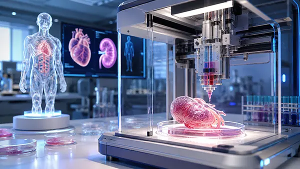
Bioprinting is often described as a future route to replacement hearts, kidneys and livers, yet its most important medical impact is already appearing in smaller and more practical forms. Researchers can arrange living cells, supportive materials and biological signals into three-dimensional tissues that resemble selected features of the human body. These constructs are being used to study disease, compare medicines and design implants around an individual patient’s anatomy. As of 2026, no fully bioprinted solid organ is available for routine transplantation, and claims that hospitals can simply “print” a new kidney remain misleading. Even so, progress in patient-derived cells, vascular engineering and tissue maturation is narrowing the distance between laboratory models and clinical treatment. The field is therefore best understood not as a single breakthrough waiting to happen, but as a sequence of advances: first better tissue models, then repair patches and small implants, followed eventually by larger and more demanding biological replacements.
From Medical Scan to Living Tissue: How Bioprinting Works
Conventional 3D printing builds an object from plastic, metal, ceramic or another non-living material. Bioprinting follows the same layer-by-layer principle but works with far more delicate ingredients. A typical “bioink” combines living cells with a water-rich gel that protects them during printing and holds them in position afterwards. The gel may imitate parts of the extracellular matrix, the natural support network surrounding cells in the body. Depending on the intended tissue, scientists can also add proteins, minerals or growth signals that encourage cells to attach, multiply and develop the required behaviour. The printer must place these ingredients accurately without exposing the cells to damaging pressure, heat or light. This is why a successful construct depends on much more than its external shape: the cells must remain alive, communicate with one another and gradually organise into functioning tissue.
The process usually begins with medical imaging or a carefully designed digital model. A scan can define the shape of a patient’s missing cartilage, bone defect or damaged tissue, while laboratory data determine which cell types and materials are needed. Cells are then expanded to obtain a sufficient number, mixed into one or more bioinks and loaded into separate printer cartridges. During printing, the machine follows the digital design and deposits each material in a planned position. Some systems push bioink through a fine nozzle, others release droplets, and light-based methods solidify selected regions of a photosensitive gel. Once printing is complete, the construct normally enters a controlled chamber or bioreactor where oxygen, nutrients, temperature and mechanical stimulation support further development.
Printing is therefore only one stage in a longer manufacturing process. A newly printed cardiac patch, for example, may contain heart muscle cells but still lack the strength, rhythm and organised structure expected of adult heart tissue. Bone constructs may require mineral development, while cartilage must gain enough resilience to withstand repeated pressure. Researchers assess cell survival, tissue shape, mechanical properties, electrical activity and biochemical function before considering implantation. They also need reliable methods that produce comparable results each time. A design that works once in a research laboratory is not automatically suitable for medical use; clinicians and regulators need evidence that every construct meets predefined standards and remains safe after implantation. Bioprinting brings precision to cell placement, but biology still needs time to complete much of the work.
Why a Patient’s Own Cells Matter
Personalisation begins with the cell source. In some cases, doctors can collect mature cells from a patient, expand them and use them to create replacement tissue of the same general type. Another approach uses induced pluripotent stem cells, often shortened to iPSCs. Scientists make these by reprogramming adult cells, such as skin or blood cells, into a stem-cell-like state. The cells can then be guided towards heart, liver, nerve or other specialised lineages. This method can provide large numbers of cells carrying the patient’s genetic background. It also makes it possible to create tissues for people whose disease cannot be reproduced accurately in standard laboratory cell lines. The result is not an exact miniature copy of the patient, but it can reflect important biological traits that influence disease and treatment response.
Using a patient’s own cells could reduce the likelihood of immune rejection, one of the major problems in transplantation, but it does not guarantee complete compatibility. Reprogrammed or extensively cultured cells may change during preparation, and the supporting gel, added proteins or manufacturing residues can provoke an immune response. Some diseases are caused by inherited variants, so tissue made from uncorrected patient cells may carry the same underlying defect. Researchers must also check that stem-cell-derived products do not contain immature cells capable of uncontrolled growth. For these reasons, personalised tissue production requires genetic checks, contamination testing, confirmation of cell identity and careful monitoring after treatment. Autologous cells are an important advantage, not a substitute for safety assessment.
The nearer-term benefit is the creation of patient-specific tissue models for selecting or developing medicines. A small bioprinted liver, tumour or cardiac tissue sample can be exposed to several treatments while the patient avoids unnecessary risk. These models can reveal whether a drug damages heart cells, whether a cancer resists a particular therapy or whether a genetic condition changes tissue behaviour. They may also provide more relevant evidence than flat cell cultures, because cells in a three-dimensional construct interact with neighbours and surrounding material in a more realistic way. In 2026, this research use is further advanced than whole-organ replacement. It is already shaping drug discovery programmes and offers a practical route towards personalised medicine even before transplantable organs become available.
What Laboratories Can Produce in 2026
The term “lab-grown organ” covers products at very different stages of development. Laboratories can produce thin skin-like tissues, cartilage constructs, bone scaffolds containing cells, vascular tubes, cardiac patches and liver microtissues. They can also bioprint organoids, which are small self-organising cell structures that reproduce selected features of an organ rather than its full size or complete function. These tissues are valuable because they can be designed with consistent dimensions and with different cell types placed in planned locations. Skin and cartilage are comparatively accessible targets because they do not require the dense internal blood supply of a liver or kidney. Even here, however, researchers must reproduce several layers, mechanical properties and long-term integration with the patient’s own tissue.
One significant clinical milestone is AuriNovo, a patient-specific biological construct studied for reconstruction of the external ear in people born with microtia. The registered US study describes a collagen-based scaffold containing the patient’s own cartilage cells and printed to match the shape of the opposite ear. An external ear is not a life-sustaining internal organ, but the work demonstrates several principles needed for personalised regenerative treatment: collection of a patient’s cells, controlled expansion, anatomical design, sterile manufacturing and surgical implantation of a living construct. It also shows why early clinical uses are likely to involve tissues with a relatively simple blood supply before researchers attempt a heart, liver or kidney.
A separate registered study in South Korea is assessing a patient-specific bioprinted tracheal construct made with hydrogels and cells obtained from nasal tissue and cartilage. The protocol is an early feasibility investigation designed for one participant, with safety and airway function monitored by imaging, endoscopy and laboratory tests. This is a highly limited experiment rather than proof that bioprinted airways are ready for general treatment. Its importance lies in the move from laboratory testing towards carefully supervised human use. Together, such studies suggest that clinical translation will occur tissue by tissue, with narrow indications, small participant groups and long follow-up periods. Progress should therefore be judged by verified safety and sustained function, not by the visual resemblance of a printed construct to a natural organ.
Why Full Solid Organs Remain Difficult
The greatest obstacle is blood supply. Most living cells must remain close to tiny vessels that deliver oxygen and nutrients and remove waste. A thin piece of tissue can receive some support by diffusion, but cells deep inside a full-sized organ quickly become deprived unless the construct contains a connected, perfusable vascular network. This network must include larger channels as well as capillary-scale branches and must link rapidly with the recipient’s circulation. Research published in 2026 demonstrated printed channels below ten micrometres and showed that endothelial cells could form continuous linings inside them. That is meaningful progress, yet a natural organ contains an enormous, adaptive vascular tree that responds to pressure, injury and changing metabolic demand. Reproducing that entire system reliably remains a major challenge.
A solid organ is also a community of specialised cell types arranged across several scales. The kidney contains filtering units, vessels, collecting structures and supporting cells organised with microscopic precision. The liver must process nutrients, neutralise harmful substances, produce proteins and manage bile flow. The heart needs aligned muscle fibres, electrical coordination, valves and vessels capable of working without interruption. Printing the outline of these organs is comparatively straightforward; reproducing their internal organisation and continuous function is not. Cells also need to mature after printing, and many stem-cell-derived cells initially behave more like foetal than adult tissue. A construct may perform one useful task in the laboratory while still falling far short of the combined workload required inside the human body.
Scale creates further problems in production and quality control. A small tissue model can be examined under a microscope and manufactured in a multiwell plate, whereas a transplant-sized organ may contain billions of cells distributed through a thick structure. Scientists need to confirm that the correct cells are present in the correct places, that channels remain open, that no region is dying and that the tissue responds normally under stress. They must also control the rate at which temporary materials break down as natural tissue develops. Storage and transport are difficult because a living construct cannot always be frozen or kept on a shelf like a conventional device. Each additional cell type, material and manufacturing stage increases the number of variables that must be measured and controlled.
How Bioprinting Could Change Personalised Medicine
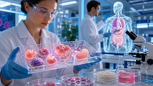
The most immediate change may occur before a patient enters an operating theatre. Bioprinted tissues can help researchers test medicines on human-like models at an early stage, identify toxic effects and compare responses between healthy and diseased cells. The US National Center for Advancing Translational Sciences is developing printed models of tissues including skin, liver, lung, retina, brain, placenta, skeletal muscle and cardiac muscle for disease research and drug screening. In 2026, its work also includes a bioprinted skin model used in research on treatments for herpes simplex virus. Such models do not replace every animal study or clinical trial, but they can improve the evidence used to decide which treatments deserve further development and which may be unsafe or ineffective.
Bioprinting can also support more tailored surgery. A construct based on a patient’s scan can fit an irregular defect more closely than a standard implant, which may be useful in cartilage repair, facial reconstruction, bone regeneration or wound treatment. Surgeons could eventually receive tissue with the required geometry, cell composition and degradation rate rather than adapting a mass-produced implant during an operation. In situ bioprinting takes the idea further by depositing cells and materials directly into a wound or defect, although most internal applications remain experimental. The goal is not merely cosmetic accuracy. A better anatomical fit can influence load distribution, tissue contact and healing, while planned placement of cells and biological signals may encourage the patient’s own tissue to integrate with the repair.
For transplantation, the long-term promise is a supply of organs designed around the recipient rather than limited by donor availability. Patient-derived cells could lower some rejection risks and may reduce dependence on lifelong immunosuppressive treatment, although this benefit has not yet been demonstrated for complete bioprinted solid organs. Personalised design could also account for body size, vessel arrangement and previous surgery. A realistic development path will probably involve partial solutions first: liver tissue that temporarily supports a failing organ, a cardiac patch that repairs damaged muscle, a kidney component that restores one function or a vascularised graft that replaces a limited region. These products could deliver meaningful clinical value without reproducing every feature of a natural organ from the beginning.
Safety, Regulation and the Realistic Road Ahead
Every bioprinted implant combines risks associated with cells, biomaterials, manufacturing and surgery. Cells can become contaminated, change genetically, mature unevenly or behave differently after implantation. Hydrogels and temporary scaffolds must support the tissue long enough, then degrade without releasing harmful substances or causing damaging inflammation. Printed blood channels must not leak, collapse or trigger clotting. An implant that appears functional for several weeks may fail after months of mechanical strain or immune activity. Safety testing must therefore cover sterility, cell identity, structural strength, biological function, degradation and the possibility of tumour formation. Long-term follow-up is especially important because living products can continue to change after they enter the body.
Regulation is complicated by personalisation. Traditional medicines are manufactured in large batches and tested against a fixed specification, while a patient-specific tissue may differ in shape, cell source and production schedule for every recipient. Authorities still need evidence that the process is controlled and that those differences remain within safe limits. Manufacturers must record where cells came from, how they were expanded, which materials were used and whether the final construct met release criteria. Hospitals may also need specialised facilities and trained staff if production takes place near the point of care. Clear rules for consent, cell ownership, genetic data, cost and access will be necessary so that personalised treatment does not create avoidable ethical or social inequalities.
The position in 2026 is promising but measured. Bioprinted tissue models are already useful in research, and early human studies of personalised cartilage and airway constructs show that selected implants are entering clinical evaluation. At the same time, fully functional printed hearts, kidneys, livers and lungs remain research goals rather than available treatments. The next credible advances are likely to involve better vascularised tissues, more mature patient-derived cells, automated quality checks and products designed to repair part of an organ. No reliable date can be given for routine whole-organ bioprinting, because progress depends on biological performance, manufacturing consistency and long-term safety rather than printing speed alone. The field is moving personalised medicine forward, but it will do so through carefully tested stages rather than one dramatic leap.